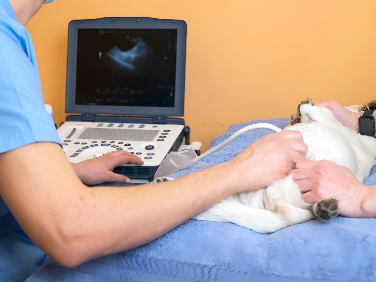
Pet Ultrasound
At PetSmart Veterinary Services in Garner, NC, we base our individualized care on thorough and accurate diagnostics like our pet ultrasound services.
What is Pet Ultrasound?
Pet ultrasound is a diagnostic imaging technique that uses sound waves to create detailed images of the internal organs and structures of your pet’s body. Unlike X-Rays, which provide images of bones and dense tissues, ultrasounds are excellent for examining soft tissues, such as the liver, kidneys, heart, and abdominal organs. This makes it an essential tool for diagnosing and monitoring a wide range of conditions.
Key Services Offered
At PetSmart Veterinary Services in Garner, NC, our comprehensive pet ultrasound services include:
- Abdominal Ultrasounds: To diagnose and monitor conditions affecting the liver, kidneys, intestines, bladder, and other abdominal organs.
- Cardiac Ultrasounds (Echocardiograms): To evaluate heart function and diagnose heart disease.
- Pregnancy Monitoring: To confirm pregnancy and monitor the development of fetuses.
- Soft Tissue Imaging: To examine masses, cysts, and other soft tissue structures.

When is Pet Ultrasound Necessary?
Abdominal Issues
Ultrasounds are commonly used to investigate abdominal issues. If your pet is experiencing symptoms such as vomiting, diarrhea, or unexplained weight loss, an ultrasound can help identify the cause. It can reveal conditions like liver disease, kidney stones, and gastrointestinal blockages.
Cardiac Evaluation
For pets with suspected heart conditions, ultrasounds (or echocardiograms) are crucial. They provide detailed images of the heart’s structure and function, helping diagnose heart disease, valve issues, and other cardiac problems.
Reproductive Health
Ultrasounds are also used to monitor pregnancy and reproductive health in pets. They can confirm pregnancy, estimate the number of fetuses, and monitor fetal development, ensuring the health of both the mother and the offspring.
Monitoring Chronic Conditions
For pets with chronic conditions such as liver or kidney disease, regular ultrasounds can monitor the progression of the disease and assess the effectiveness of treatments. This allows our veterinarians to make necessary adjustments to your pet’s care plan.
The Ultrasound Process
- Preparation: Preparation for a pet ultrasound typically involves fasting your pet for several hours before the procedure. This helps reduce the amount of gas in the intestines, which can interfere with the imaging. Our team will provide specific instructions based on your pet’s needs.
- The Procedure: During the ultrasound, your pet will lie comfortably on a padded table. A small amount of fur may be shaved from the area being examined to allow better contact with the ultrasound probe. A special gel is applied to the skin to facilitate the transmission of sound waves. The veterinarian will gently move the probe over the area, capturing real-time images on a monitor.
- After the Ultrasound: After the procedure, your pet can return to normal activities immediately. The veterinarian will review the images and discuss the findings with you, explaining any diagnosed conditions and recommending appropriate treatments or further testing if necessary.
